Prophase Under Microscope
Microtubules attach at the kinetochores and the chromosomes begin moving. If fully stretched out some DNA may be nearly a centimeter long much too large for a cell nucleolus.
:max_bytes(150000):strip_icc()/interphase-58e3d4a45f9b58ef7e071ea0.jpg)
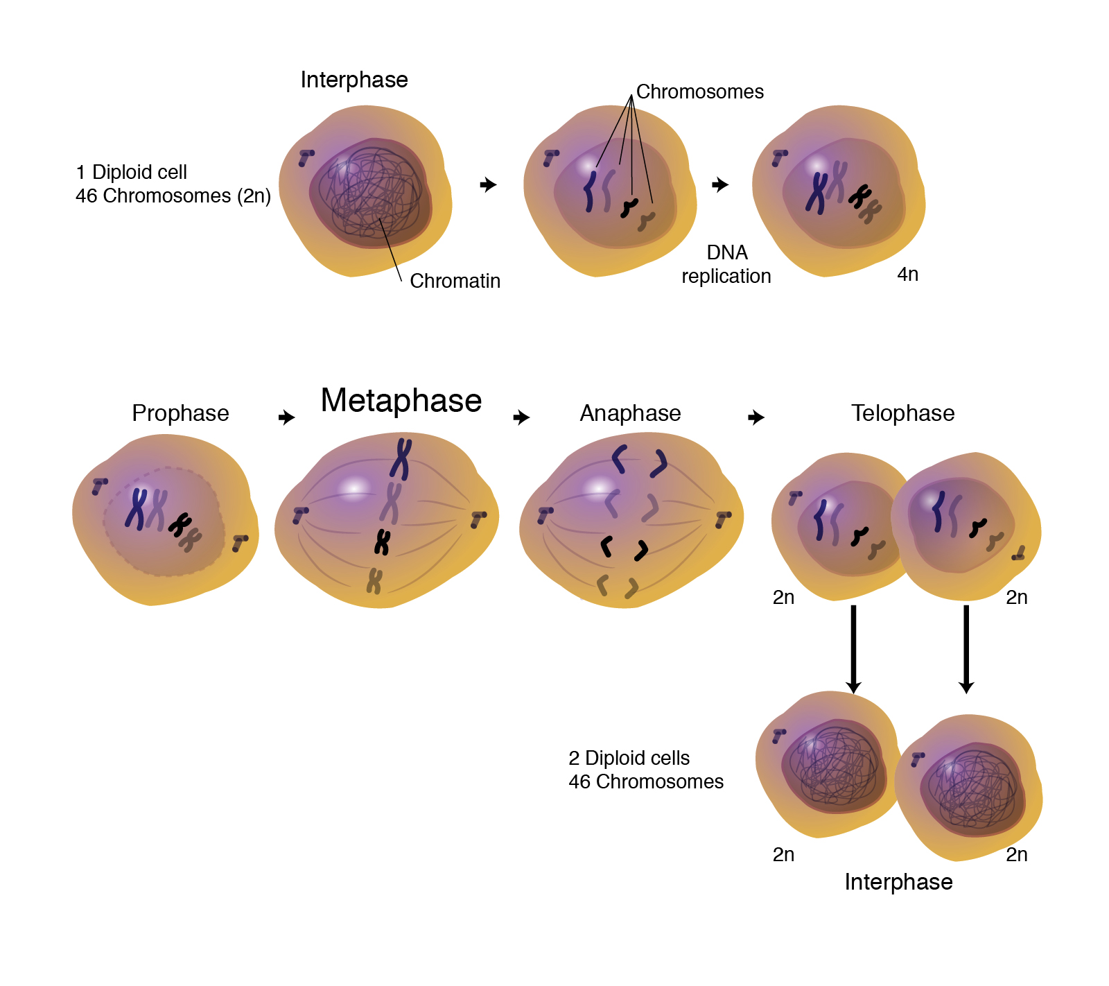
The Stages Of Mitosis And Cell Division
Can You See Chromosomes With A Light Microscope During Meiosis I Know It S Possible When The Chromosomes Condense During Mitosis But I Don T Know About During Meiosis Quora

Mitotic Cell Division 1 Objectives 2 Learn Preparing
In Mitosis Prophase Chromatin in the nucleus begins to condense and becomes visible in the light microscope as chromosomes.

Prophase under microscope. The 4 Phases of Mitosis. You may use your textbook and class notes to help you identify the stages of mitosis as seen under the microscope. The student will correctly identify and draw four stages of mitosis using microscope slide images of onion root tips and whitefish blastulae.
When we first see chiasmata under a microscope we know that _____. Chromatin in the nucleus begins to condense and becomes visible in the light microscope as chromosomes. Nuclear Envelope breaks down.
The nuclear envelope breaks down. Centrioles begin moving to opposite ends of the cell and fibers extend from the centromeres. A karyotype is a technique that allows researchers to visualize the chromosomes under the microscope with the help of proper extraction and staining techniques.
In late prophase I homologous chromosomes also called bivalent chromosomes or bivalents pair laterally or side-by-side. Some fibers cross the cell to form the mitotic spindle. Proteins attach to the centromeres creating the kinetochores.
The nucleolus is a dark spot in the nucleus that contains RNA and proteins responsible for creating ribosomes. During prophase I chromosomal condensation allows chromosomes to be viewed under the microscope. The chiasmata finally arrive at the end of the chromatid arms of the chromosomes finalizing terminalization.
Mitosis is responsible for a single cell a fertilized human embryo developing into a human body with. Most of the time DNA is tightly coiled and structured around proteins called histones. _____ may be employed to use computer algorithms to.
From Ancient Greek μείωσις meiosis lessening because it is a reductional division is a special type of cell division of germ cells in sexually-reproducing organisms used to produce the gametes such as sperm or egg cellsIt involves two rounds of division that ultimately result in four cells with only one copy of each chromosome. This packaged form is known as chromatin. M is the actual period of cell division consisting of prophase metaphase anaphase telophase and cytokinesis.
This means that during prophase I the chromosomes condense becoming thicker and shorter. It takes place after the chromatids have condensed and the sister chromatids are bivalent of tetrad as visualized under a microscope. The modern definition of a chromosome now includes the function of heredity and the chemical composition.
Other key events are. Chromatin in the nucleus begins to condense and becomes visible under the light microscope as chromosomes each with two chromatids that are held together at the centromere. Prophase I is occurring Homologous pairs of chromosomes align opposite to each other at the equator of a cell during ________.
During this stage the chromosomes super coil condense and become visible for first time during the cell cycle. Additionally well mention three other intermediary stages interphase prometaphase and cytokinesis that play a role in mitosis. Prophase - The chromosomes supercoil and become visible under a light microscope.
Cytokinesis interphase anaphase prophase metaphase. At this first stage of Prophase I of meiosis I chromosomes are visible under electron microscopy and look like a string of beads where the beads are referred to as nucleosomes. What Happens During Prophase I.
Centrioles begin moving to opposite ends of the cell and fibers extend from the centromeres. Karyokinesis or mitosis is divided into five stagesprophase prometaphase metaphase anaphase and telophase. The first phase of mitosis is known as the prophase where the nuclear chromatin starts to become organized and condenses into thick strands that eventually become chromosomes.
The chromosomes assume their classic X shape - two sister chromatids joined in the middle at the centromere. Diakinesis is the final step of Prophase 1 and is the termination of the condensing of the chromosomes this allows the chiasmata and bivalent structure to be seen more clearly under an electron microscope. By the time prophase I of meiosis begins the chromosomes within the cell have duplicated and prepared for cellular division.
Chromatin in the nucleus begins to condense and becomes visible in the light microscope as chromosomes. Each chromosome is composed of two sister chromatids containing identical genetic information. The copied chromosomes condense into X-shaped structures that can be easily seen under a microscope.
Under the microscope in its extended form chromatin looks like beads on a string. It is therefore packaged using special proteins. The four stages of mitosis are known as prophase metaphase anaphase telophase.
Diakinesis-The fifth and final phase of prophase I. Chromosomes were first named by cytologists viewing dividing cells through a microscope. The beads are called nucleosomes.
Centrioles begin moving to opposite ends of the cell. Chromosomes first visible - thanks to supercoiling - under a light microscope at Prophase. Another component of the nucleus the nucleolus disappears during prophase.
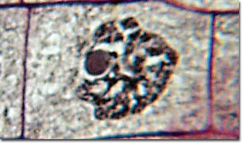
Under a microscope this stage can be seen as a darkening of different places in the nucleus. The nuclear membrane dissolves marking the beginning of prometaphase. The main occurrences in prophase are the condensation of the chromatin reticulum and the disappearance of the nucleolus.
If you were looking under the compound light microscope at an onion root tip in what stage of the cell cycle would the majority of the cells be. The chromosomes pair up so that both copies of chromosome 1 are together both copies of chromosome 2 are together and so on. B _____ is the study of the structure and function of cellular proteins and their interactions.
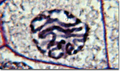
Each nucleosome is composed of DNA wrapped around eight proteins called histones. Prophase under a microscope during prophase the molecules of dna condense becoming shorter and thicker until they take on the traditional x shaped appearance. The slides below show longitudinal sections of allium onion root tip.
Prophase Metaphase Anaphase Telophase. During prophase the cytoskeleton composed of cytoplasmic microtubules begins to disassemble and the main component of the mitotic apparatus the mitotic spindle begins to form outside the nucleus at. The pictures at the bottom were taken by fluorescence microscopy hence the black background of cells artificially stained by fluorescent dyes.
It sets up the cell for metaphase. Meiosis m aɪ ˈ oʊ s ɪ s. The chromosomes are at their most condensed form during diakinesis.
This makes the chromosomes visible under a light microscope. So what are the stages of mitosis. At this time they are said to be in synapsis.
Prophase from Ancient Greek προ- before and φάσις phásis appearance is the first stage of cell division in both mitosis and meiosisBeginning after interphase DNA has already been replicated when the cell enters prophase. Blue fluorescence indicates DNA chromosomes and green fluorescence indicates microtubules spindle apparatus.
Molecular Expressions Photo Gallery Mitosis
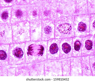
Mitosis Onion Cells Root Meristem Central Stock Photo Edit Now 159810452
Mitosis Stages Of The Lily Microbehunter Microscopy
Metaphase
Molecular Expressions Photo Gallery Mitosis
Late Prophase Of Mitosis Lm Stock Image P673 0071 Science Photo Library

Solved Observing Mitosis And Cytokineses In Plant Cells The Root Tip Of Plants Is An Area Of Rapid Cell Division Using The Virtual Microscope You Course Hero
Mic Uk Onion Root Mitosis

0 Response to "Prophase Under Microscope"
Post a Comment